
Screening for breast cancer refers to medical processes designed to detect cancerous changes in breast tissue as early as possible, often before symptoms develop. These strategies utilize various imaging and sampling technologies to find potential tumors and assess them for risk and characteristics. The underlying aim is to deliver objective, evidence-informed information that can guide later decision-making regarding further diagnostic steps or interventions. Early identification of breast abnormalities may help improve the range of management options available to individuals.
Modern breast cancer screening focuses on applying specialized imaging and laboratory techniques to identify subtle differences in breast tissue. Routine screening is generally offered to certain populations based on age or individual risk factors, with exact protocols differing by health authority and current evidence. Diagnostic follow-up may be recommended if screening identifies areas of concern. Methods in common use have varying levels of sensitivity and specificity, and each has unique advantages and limitations. The choice of screening approach typically reflects an individual’s health profile, medical history, and access to care.
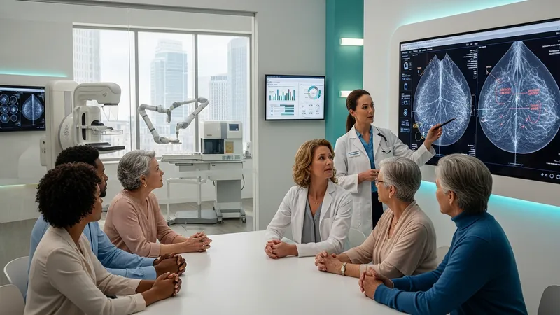
Mammography is frequently regarded as the foundational imaging method for breast cancer screening. It has been studied extensively for its ability to identify early-stage changes, such as microcalcifications. However, mammography's sensitivity may decrease in individuals with denser breast tissue, which is why alternative or supplemental imaging methods can be considered for certain populations.
Ultrasound is not typically used as a primary screening tool, but it plays an important role in further characterizing findings from mammograms, particularly in evaluating cystic versus solid masses. It is non-invasive and does not use ionizing radiation, making it suitable for supplemental use in cases where mammographic images are inconclusive or additional clarity is needed.
Biopsy does not function as a screening test but serves as the definitive diagnostic step after imaging identifies a potentially concerning abnormality. The tissue obtained through biopsy is examined by pathologists to confirm if cancer is present and to detail the type and grade. The results from biopsies inform further management planning and are often required for a firm diagnosis.
The effectiveness of each method can vary by individual factors such as age, breast density, and specific risk profiles. Screening strategies are sometimes adapted based on evolving evidence or technological advances, which can impact sensitivity, specificity, and user experience. Individuals are generally informed of the potential benefits and limitations associated with each approach.
Overall, the primary objective of breast cancer screening and diagnostic techniques is to enable earlier recognition of abnormal tissue changes, which may support more timely and informed healthcare decisions. The next sections examine practical components and considerations in more detail.
Breast cancer detection relies upon several approaches that balance sensitivity, specificity, accessibility, and patient comfort. Mammography, as a widely established tool, typically serves as the primary screening method for average-risk populations. Its implementation in routine screening programs has contributed to the early identification of non-palpable lesions in large population groups. The technique is especially effective in individuals aged 50 and older, although it may be offered starting at earlier ages based on individual circumstances and emerging guidelines.
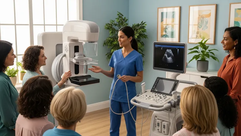
Ultrasound is often incorporated as a secondary imaging modality rather than a universal screening tool. Its particular strength lies in its use with denser breast tissue or younger individuals for whom mammography may yield less reliable images. Ultrasound can distinguish between solid and fluid-filled lesions, thereby narrowing down follow-up options and preventing unnecessary invasive procedures in certain cases. It is generally non-invasive and does not expose recipients to ionizing radiation, making it amenable to repeated use in diagnostic workups.
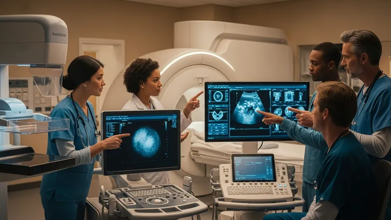
Biopsies are conducted when imaging methods reveal a mass or abnormality that cannot be conclusively classified as benign or malignant. The process involves extracting a minute tissue sample, often with a needle guided by imaging, minimizing invasiveness. The specific technique—such as core needle, fine-needle aspiration, or surgical biopsy—may vary based on the lesion’s location, size, and initial imaging results. Pathological analysis of biopsy material provides the definitive assessment for malignancy and offers details about the tumor’s properties.
The decision on which combination of screening or diagnostic tests to use frequently depends on a balance of clinical findings, risk factors, and guideline-based recommendations. There is no universally optimal sequence; instead, healthcare providers may tailor protocols to fit individual needs while taking into account the strengths and limitations of each available method. This multifaceted approach underscores the complex, evolving landscape of breast cancer detection.
The outcomes of breast cancer screening are influenced by several factors, including age, breast density, genetic risks, and personal or family medical history. For instance, individuals with dense breast tissue may experience lower sensitivity with mammography, making it more likely for additional tests such as ultrasound or magnetic resonance imaging to be recommended. Screening protocols often reflect current research into which factors most impact test accuracy and interpretation.
Technological advancements in imaging have gradually improved the detection rates of small or early-stage lesions, but with increased detection comes the possibility of higher false-positive rates. This can result in anxiety, additional testing, and, in rare cases, biopsies of ultimately benign areas. Balancing the benefits of early detection with the potential drawbacks of overdiagnosis or unnecessary procedures is a core consideration in modern screening programs.
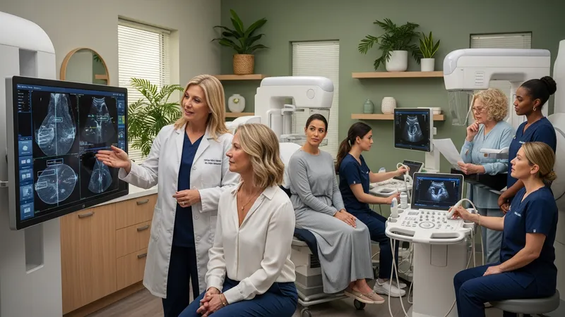
Another factor is access to high-quality screening and diagnostic services, which may vary depending on location and healthcare system resources. Regular quality assurance and accreditation standards are designed to enhance imaging consistency and interpretation. Some programs include double-reading of mammograms or the use of computer-aided detection software to bolster the identification of subtle findings, although final diagnosis always relies on human expertise.
The communication of results is also an important factor in the screening process. Individuals who undergo screening are typically provided with an explanation of their results, guidance for next steps if further evaluation is advised, and information regarding possible interval follow-up. Clear reporting practices help ensure that participants have the necessary context to understand their screening outcomes and subsequent care pathways.
Screening techniques are not without limitations, and these challenges are important to recognize when interpreting results or making decisions about further testing. Mammography, while effective for many, may yield false-negative or false-positive results, particularly in younger individuals or those with denser tissue. Image artifacts, technical variability, or overlapping structures can all contribute to limitations in image interpretation. This sometimes prompts the use of alternative imaging, such as ultrasound or, when indicated, magnetic resonance imaging.
Ultrasound, as a supplementary screening option, can be limited by operator dependence and variability in resolution based on equipment quality. Although useful in distinguishing between cystic and solid lesions, ultrasound does not always offer sufficient detail to rule out all types of abnormalities. It is generally not used as the sole screening modality for breast cancer due to these restrictions, instead working best as part of a diagnostic sequence following initial imaging findings.
Biopsy procedures introduce their own set of considerations, such as minor risk of discomfort, infection, or bleeding at the sample site. The procedural technique chosen is dependent on abnormality size, depth, and location, with an emphasis on minimizing invasiveness where possible. Misclassification or non-representative sampling, while rare, can potentially impact the accuracy of the diagnosis and lead to additional procedures.
Overall, the interpretation of results from these screening and diagnostic methods requires integration with individual risk profiles, past medical history, and evolving guidelines. As new data and technologies continue to emerge, best practices in screening protocols may adapt to maximize benefit and reduce potential harms. Ongoing research supports the refinement of protocols for different populations and risk groups.
Emerging advancements in breast cancer screening include the development of digital mammography and tomosynthesis (3D mammography), which may improve detection rates of small lesions in certain populations. Digital systems can offer enhanced image storage, transmission, and analysis opportunities, including the potential for computer-aided detection algorithms to assist radiologists. Ongoing research is examining whether these newer technologies lead to consistently improved outcomes across different age and risk groups.

Molecular imaging and the integration of genetic information are also being explored as ways to tailor screening and risk assessment strategies. For example, certain gene mutations may prompt more frequent or earlier use of imaging even when physical symptoms are absent. Researchers are evaluating new biomarkers and imaging tracers that may improve specificity, although such approaches remain primarily experimental and are not yet widely implemented in routine practice.
Artificial intelligence (AI) is increasingly being integrated into breast cancer screening protocols, primarily to assist with image analysis, reduce interpretation variability, and prioritize cases for review by specialists. Clinical studies are ongoing to determine the most effective means of using AI as a supportive tool while ensuring human oversight remains central to diagnostic decisions.
In summary, breast cancer screening continues to adapt in step with technological and scientific advances. The evolution of imaging modalities, diagnostic tools, and individualized risk assessment is likely to shape screening practices going forward. Maintaining awareness of current and emerging strategies may support informed dialogue between individuals and healthcare providers regarding the detection and assessment of breast abnormalities.