
Artificial intelligence (AI) encompasses a range of computational technologies that allow machines to process information, recognize patterns, and provide analytical support across sectors. Within the context of breast cancer, AI is most frequently applied to the analysis of complex imaging data and scientific literature. These systems rely on training algorithms to identify features in medical images and datasets that may otherwise be difficult for humans to interpret due to scale or complexity. The practical use of AI in this field centers around aiding healthcare professionals in managing extensive information, optimizing their workflows, and providing secondary analysis.
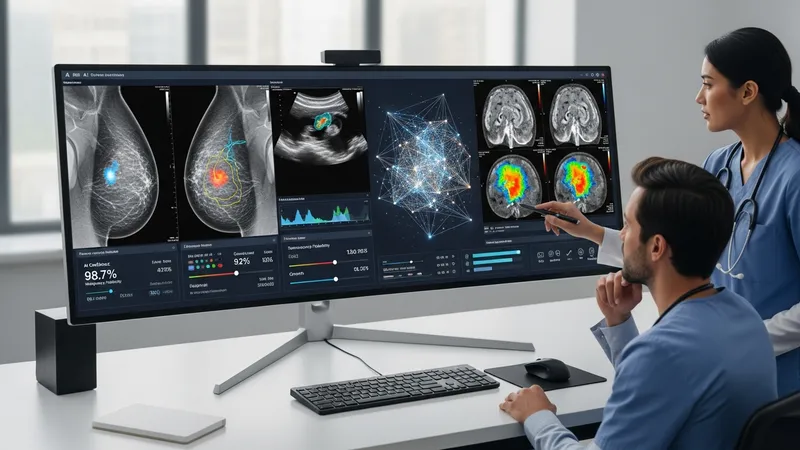
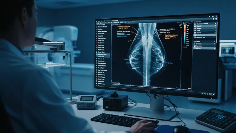
AI’s integration into breast cancer diagnostics often involves the use of software that can assess mammograms, ultrasound images, or MRI scans for notable characteristics. Deep learning algorithms, particularly convolutional neural networks, can be tailored to detect differences in tissue structure or density. In parallel, natural language processing is sometimes deployed to extract or synthesize key findings from research publications and medical records, supporting evidence-based practices. These approaches are developed as assistive tools, intended to enhance established diagnostic procedures under regulated oversight.
The development of AI in breast cancer applications is influenced by access to high-quality, annotated datasets. These datasets allow machine learning models to learn from a variety of cases, which typically improves the system’s ability to recognize relevant image features. Research initiatives often partner with hospitals or medical research institutions to obtain diverse samples, ensuring the models are exposed to a realistic range of imaging scenarios encountered in clinical practice.
AI tools in breast cancer are frequently assessed for their performance by comparing their analytical outputs to those of expert radiologists. These comparative studies may use metrics such as sensitivity and specificity to evaluate how well the AI identifies image characteristics in relation to established standards. This process is conducted under controlled conditions, typically as part of validation studies or pilot programs within healthcare networks, rather than as standalone diagnostic systems.
Emerging applications of AI extend beyond imaging to data management and workflow optimization. For example, some platforms are designed to automatically sort and organize imaging records, summarize case histories, or flag abnormalities for follow-up investigation. While the primary focus remains on assisting clinical professionals, there is also research interest in improving efficiency and reducing administrative burdens through automation of repetitive tasks.
The potential of AI in breast cancer diagnostics is closely monitored by regulatory bodies and professional associations. New tools may be subject to ongoing evaluation to ensure accuracy, data privacy, and integration with existing practices. As the landscape continues to evolve, understanding the application areas and collaborative development frameworks can provide valuable context. The next sections examine practical components and considerations in more detail.
High-quality data is foundational to the success of AI applications in breast cancer diagnostics. Models are typically trained on large, annotated image datasets, with each image labeled according to expert findings. This allows the AI system to learn the visual features that may indicate various tissue patterns. The inclusion of diverse demographic backgrounds and imaging equipment settings further enhances generalizability, ensuring the algorithms remain applicable in a range of clinical environments.
When developing AI for breast cancer analysis, data sources often comprise digitized mammograms from screening programs or hospital archives. Rigorous anonymization protocols are applied to protect patient confidentiality. Institutions may use frameworks such as the Digital Imaging and Communications in Medicine (DICOM) standard to facilitate consistent image formatting, making it easier to integrate data from multiple sites for training purposes.
AI developers may employ methods like cross-validation and iterative retraining to test and refine their models before deployment. Validation datasets, separated from the initial training set, allow for an objective assessment of how the model performs on unseen cases. Studies have found that such approaches may reduce overfitting and improve reliability when models are eventually applied in clinical settings.
Data imbalance is a commonly discussed challenge in this area. For example, rare presentations might be underrepresented in available datasets, potentially influencing model output. Strategies for addressing these gaps include data augmentation, where modifications to existing images create synthetic variations, and targeted collection efforts focused on acquiring more examples of uncommon scenarios.
Convolutional neural networks (CNNs) are among the most widely used techniques in AI-driven breast cancer imaging. These models automate the extraction of visual features from scans, learning to recognize subtle differences in tissue composition and structure. Their architecture, characterized by multiple algorithmic layers, can process complex patterns with high granularity, which is critical for mammography and tomosynthesis evaluation.

Another prevalent method involves the use of support vector machines (SVMs) in combination with handcrafted feature extraction. Radiomics approaches often calculate statistical descriptors—such as texture, shape, and edge features—from segmented images. These features are then classified by the AI to assign probabilities or scores related to specified clinical findings. SVMs are valued for their interpretability and relatively quick training on smaller datasets.
Beyond static image review, some AI platforms facilitate temporal image analysis, comparing scans from multiple time points to detect changes over time. By analyzing progression patterns, these techniques may assist clinicians in monitoring follow-up images, especially in patients with previous abnormal findings. Such longitudinal assessment is often used as a supportive tool alongside traditional evaluation practices.
Integration with picture archiving and communication systems (PACS) is a key consideration for AI-assisted image review. Many solutions are designed to be compatible with existing clinical IT infrastructure, streamlining the delivery of AI-processed results to radiologists. This integration can help ensure that new technologies are incorporated into standard diagnostic workflows with minimal disruption.
AI systems are increasingly utilized as workflow support tools within breast cancer diagnostic pathways. By pre-analyzing images and highlighting areas for radiologist review, these solutions can contribute to more structured prioritization of cases within clinical settings. Some platforms include functionalities to flag exams that may benefit from additional human oversight or expedited follow-up, potentially addressing common bottlenecks in imaging departments.
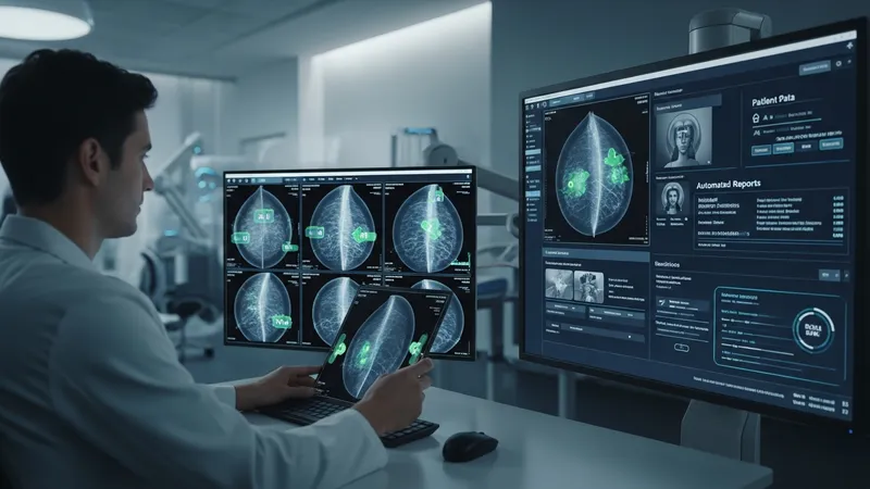
Administrative support is another area where AI can play a role. For example, automated data entry and report generation features are incorporated into certain AI-powered platforms. This may reduce manual data handling, freeing clinicians to focus on patient-facing responsibilities. Efficiency gains typically depend on the degree of interoperability with electronic health record (EHR) systems and the alignment of AI outputs with clinical documentation standards.
In multi-site healthcare networks, AI-assisted workflow tools can facilitate centralized triage of breast imaging cases. Images received from various facilities are pre-screened using standardized AI protocols, which may assist in distributing workload or balancing case review timelines. Such centralization is often governed by policy frameworks that prioritize patient privacy and data security.
It is important to note that while AI systems are designed to optimize workflow, final clinical interpretation remains the responsibility of healthcare professionals. Ongoing training and quality assurance are typically maintained to ensure that human oversight remains central in all diagnostic decisions where AI is applied.
The deployment of AI in breast cancer diagnostics is contingent upon thorough evaluation of safety, efficacy, and ethical considerations. Regulatory review processes are in place to confirm that AI tools meet clinical standards before becoming widely available. These reviews often examine model performance across diverse population groups and imaging platforms to identify any limitations or sources of variability in the results.
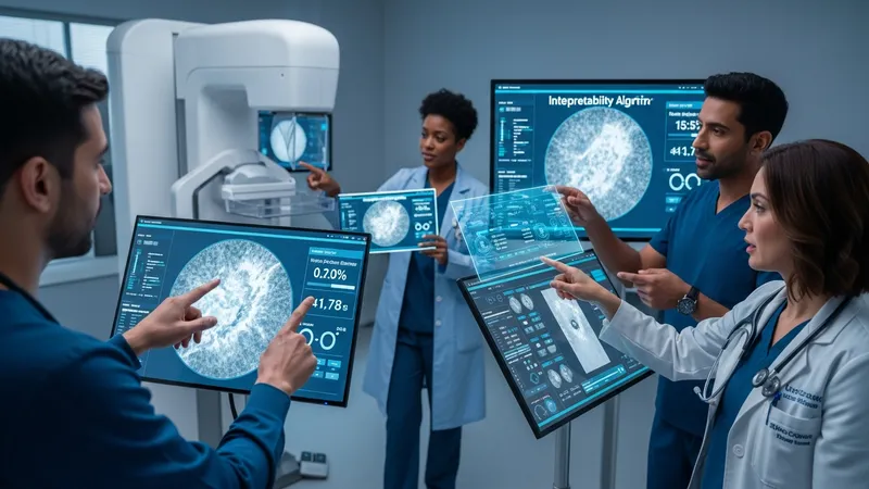
Transparency and explainability of AI models are increasingly prioritized. Clinicians and regulators generally seek insight into how algorithms reach specific conclusions based on input data. Efforts are underway to enhance the interpretability of AI outputs, ensuring that healthcare professionals can assess the rationale behind automated image assessments when incorporating them into their practice.
Data governance is key in maintaining patient trust and privacy when implementing AI systems. Institutions typically adhere to established data handling and security protocols, ensuring that patient information is protected at all stages of model training and deployment. Continuous monitoring for bias and ongoing validation are also part of quality management policies to ensure that models perform equitably.
Looking ahead, the future of AI in breast cancer diagnostics involves ongoing collaboration between technology developers, clinicians, and regulatory authorities. The careful integration of AI into established workflows is expected to remain a focus, with the intention of supporting evidence-based decision-making and maintaining high standards of patient care.